

Show Notes 12 April 2024
All Medical Miracles this Week!
Story 1: New self-powered throat patch could help people speak without vocal cords.
Source: LiveScience.com Story by Rebecca Sohn
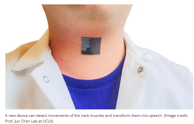
- A new, flexible device that clings to the neck can translate muscular movements into speech, enabling people to talk without using their vocal cords.
- Created by researchers at the University of California, Los Angeles, the small patch not only detects the movements of the throat associated with speech, but also harnesses that movement to generate electricity, meaning the device can be operated without a battery or being plugged in.
- The device, described in a study published March 12 in the journal Nature Communications, could theoretically help people with voice disorders caused by damaged or paralyzed vocal cords to communicate, including those recovering from throat cancer surgery.
- Lead study author Jun Chen, an assistant professor of bioengineering at the University of California, Los Angeles, says the idea for the patch arose after he’d spent several hours lecturing and felt his voice becoming tired. He began to imagine a way to solve this problem, to make it possible for a person to speak without using their vocal cords, also known as “vocal folds” [a.k.a. Vocal Cords].

Jun Chen
- How it works:
- Scientists have known that the magnetic properties of some rigid metals can be changed when they’re put under mechanical stress. An example of this is an alloy of iron and gallium, called Galfenol, whose magnetic state changes when you squeeze or deform the material. In their 2021 study, Chen and his colleagues showed the same concept could work with a soft material made of tiny magnets embedded inside thin silicon.
- Now, in their latest study, the team has harnessed this material in a patch that responds to the subtle stress placed on it by the movement of throat muscles. When a person executes the movements needed to talk, the material responds by producing electrical signals that can be translated into speech.
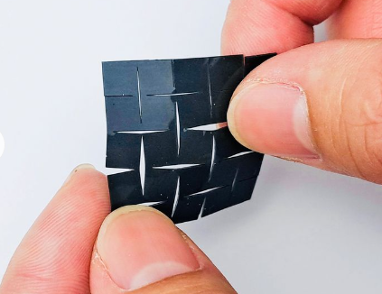
- To accomplish this, the patch is made of five very thin layers. The outer layers of the patch are made of a soft, flexible silicon material, while the middle layer, made of silicon and micromagnets, generates a magnetic field that varies with the movement of the throat muscles. The two layers surrounding it, made of coils of copper wire, translate these magnetic-field changes into electrical signals.
- These electrical signals then get fed to a machine-learning algorithm that translates the pulses into speech. To train the algorithm, each participant in the study repeated five short phrases 100 times each while the program tracked their throat movements. This taught the system to associate specific movements with a given phrase.

Story 2: A bioelectronic mesh capable of growing with cardiac tissues for comprehensive heart monitoring.
Source: MedicalXpress.com Story by Daegan Miller
Link: https://medicalxpress.com/news/2024-03-bioelectronic-mesh-capable-cardiac-tissues.html
- The heart is very sensitive to therapeutic drugs, and the pharmaceutical industry spends millions of dollars in testing to make sure that its products are safe. However, ways to effectively monitor living cardiac tissue are extremely limited.
- In part, this is because it is very risky to implant sensors in a living heart, but also because the heart is a complex kind of muscle with more than one thing that needs monitoring.
- But today’s sensors can typically only measure one characteristic at a time, and a two-sensor device that could measure both charge [meaning electrical signal] and movement would be so bulky as to impede the cardiac tissue’s function. Until now, there was no single sensor capable of measuring the heart’s dual properties of charge [or electrical signal] and movement without interfering with its functioning.
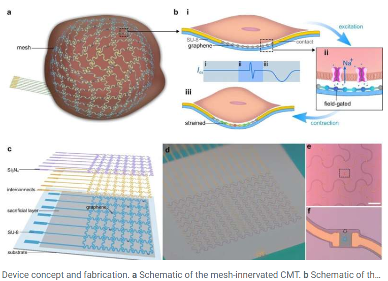
- A team of engineers led by the University of Massachusetts Amherst and including colleagues from the Massachusetts Institute of Technology (MIT) recently announced in Nature Communications that they had successfully built a [single sensor] tissue-like bioelectronic mesh system integrated with an array of atom-thin graphene sensors.
- This tissue-like bioelectronic mesh system can simultaneously measure both the electrical signal and the physical movement of cells in lab-grown human cardiac tissue.
- The new device is built of two critical components. The first is a three-dimensional cardiac microtissue (CMT) grown in a lab from human stem cells.
- The second critical component involves graphene—a pure-carbon substance only one atom thick. Graphene has a few surprising quirks to its nature that make it perfect for a cardiac sensor.
- Graphene is electrically conductive, and so it can sense the electrical charges shooting through cardiac tissue.
- It is also piezoresistive, which means that as it is stretched—for example, by the beating of a heart—its electrical resistance increases.
- In a research first, this tissue-like mesh can grow along with the cardiac cells, allowing researchers to observe how the heart’s mechanical and electrical functions change during the developmental process. The new device is a boon for those studying cardiac disease as well as those studying the potentially toxic side effects of many common drug therapies.
- One of the lead researchers noted, “No one has ever done this before. Graphene can survive in a biological environment without degrading for a very long time and not lose its conductivity, so we can monitor the cardiac microtissue across its entire maturation process.”

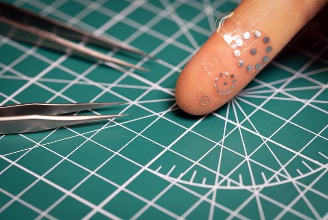
Story 3: Shape-shifting ultrasound stickers detect post-surgical complications – First-of-its-kind device ‘tags’ an organ to monitor abnormal, life-threatening fluid leaks.
Source: Northwestern University Story by Amanda Morris
- All gastrointestinal surgeries carry the risk of anastomotic leaks. If the leak is not detected early enough, the patient has a 30% chance of spending up to six months in the hospital and a 20% chance of dying.
- For example, for patients recovering from pancreatic surgery, the risks are even higher – a staggering 40-60% of patients suffer complications after pancreas-related surgeries.
- Currently, no existing methods can reliably and non-invasively detect anastomotic leaks — a life-threatening condition that occurs when gastrointestinal fluids escape the digestive system.
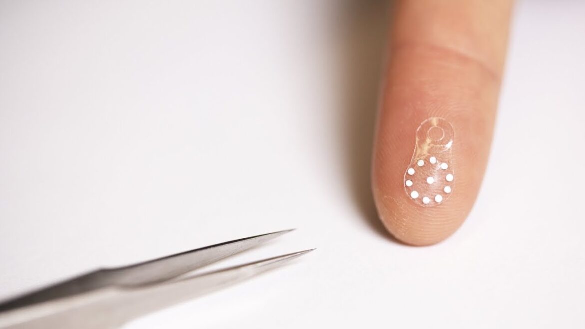
- Researchers led by Northwestern University and Washington University School of Medicine in St. Louis have developed a new, first-of-its-kind tiny sticker [comprised of a hydrogel with embedded tiny metal dots].
- The new sticker enables clinicians to monitor the health of patients’ organs and deep tissues with a simple ultrasound device.
- Here’s how it works:
- When attached to an organ, the soft, tiny sticker changes in shape in response to the body’s changing pH levels, which can serve as an early warning sign for post-surgery complications such as anastomotic leaks.
- In specific, as the hydrogel swells in response to changing pH, the metal disks move apart.
- Clinicians then can view these shape changes [reflected by the changing positions of the embedded metal dots] in real time through ultrasound imaging.
- As one of the lead researchers noted, “Because the acoustic properties of the metal disks are much different than those of the surrounding tissue, they provide very strong contrast in ultrasound images. In this way, we can essentially ‘tag’ an organ for monitoring.”
- When the patient has fully recovered, the biocompatible, bioresorbable sticker simply dissolves away — bypassing the need for surgical extraction.

Story 4: Implantable batteries can run on the body’s own oxygen.
Source: Phys.org Story by “Cell Press”
Link: https://phys.org/news/2024-03-implantable-batteries-body-oxygen.html
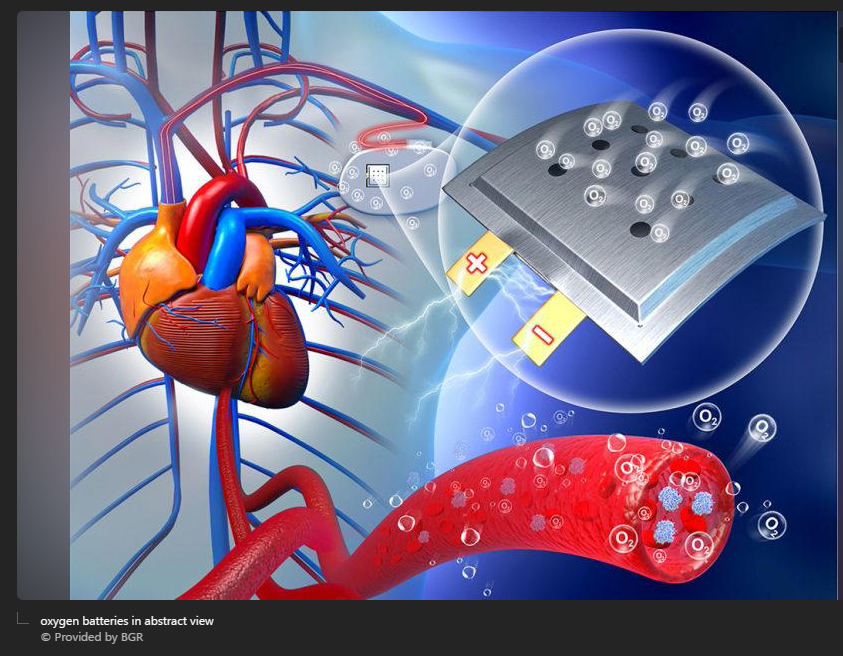
- From pacemakers to neurostimulators, implantable medical devices rely on batteries to keep the heart on beat and to dampen pain. But batteries eventually run low and require invasive surgeries to replace.
- To address these challenges, researchers at the Tianjin University of Technology in China devised an implantable battery that runs on oxygen in the body. The study, published March 27 in the journal Chem, shows that in rats the implanted proof-of-concept design can deliver stable power and is compatible with biological systems.
- To build a safe and efficient battery, the researchers made its electrodes out of a sodium-based alloy and nanoporous gold, a material with pores thousands of times smaller than a hair’s width.
- Gold has been known for its compatibility with living systems, and sodium is an essential and ubiquitous element in the human body. The electrodes undergo chemical reactions with oxygen in the body to produce electricity.
- To protect the battery, the researchers encased it within a porous polymer film that is soft and flexible.
- The researchers then implanted the battery under the skin on the backs of rats and measured its electricity output. Two weeks later, they found that the battery could produce stable voltages between 1.3 volts and 1.4 volts.
- Although the output is insufficient to power medical devices, the design shows that harnessing oxygen in the body for energy is possible. ***My comment, as this is initial research you can bet these researchers will continue to work on this technology and increase power output.
- The team also evaluated inflammatory reactions, metabolic changes, and tissue regeneration around the battery. The rats showed no apparent inflammation.

4 Honorable Mentions:
Story: Scientists invent ultra-thin, minimally invasive pacemaker controlled by light.
Source: MedicalXpress.com Story by staff

- A team of researchers with the University of Chicago has developed a wireless device, powered by light, that can be implanted to regulate cardiovascular or neural activity in the body. The featherlight membranes, thinner than a human hair, can be inserted with minimally invasive surgery and contain no moving parts.
- Published Feb. 21 in Nature, the results could help reduce complications in heart surgery and offer new horizons for future devices.
////////////////////////////////////////////////////////////////////////////////////////////////////////////////////////////////////////////
Story: New Synthetic Blood Clotting System Could Help Stop Internal Bleeding.
Source: ScienceAlert.com Story by David Nield
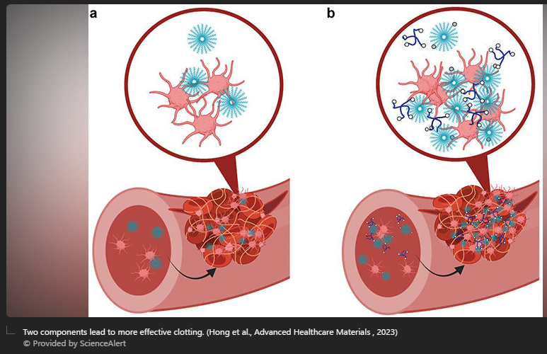
- Blood clots are one of the body’s most important natural defense systems, a mechanism for plugging internal and external gaps to keep us alive. However, in cases where the body is losing a lot of blood, the clotting process can’t keep up. This is where a new synthetic replacement could come in.
- Researchers have developed a two-component system that targets internal injuries without causing any unwanted damage of its own. The two components match the body’s platelets (cell fragments that trigger clotting) and fibrinogen (a protein that helps clots to form).
- So far, the synthetic process has only been tested on mice, but it effectively triggered the blood clotting part of the natural hemostasis reaction to wounds and proved significantly better at stopping bleeding than previous approaches.
////////////////////////////////////////////////////////////////////////////////////////////////////////////////////////////////////////////
Story: Scientists create artificial mucus to probe early tumor formation
Source: Interesting Engineering Story by Mrigakshi Dixit
Link: https://interestingengineering.com/health/scientists-create-artificial-mucus
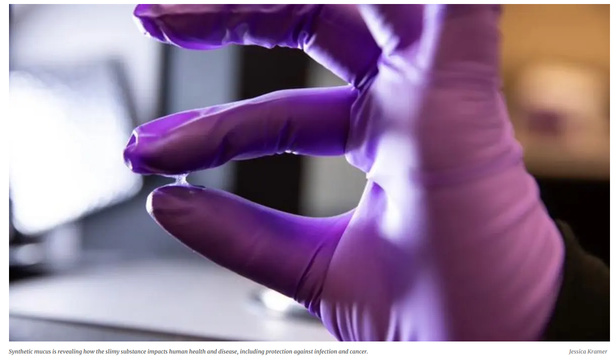
- A new study has unraveled the many roles of mucus, which is a sticky substance discharged during cold and flu season.
- The University of Utah researchers synthesized a key component of mucus called mucins. c
- These sugar-coated proteins not only govern molecular transport but also play important functions in immunology and cell behavior.
- In this new study, researchers used synthetic mucin to understand how it affects the early phases of tumor formation.
////////////////////////////////////////////////////////////////////////////////////////////////////////////////////////////////////////////
Story: Doctors can now watch spinal cord activity during surgery
Source: ScienceDaily.com University of California Riverside
Link: https://www.sciencedaily.com/releases/2024/03/240307110756.htm
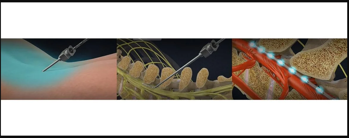
- With technology developed at UC Riverside, scientists can, for the first time, make high resolution images of the human spinal cord during surgery. The advancement could help bring real relief to millions suffering chronic back pain.
Leave a Reply